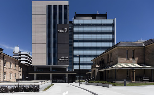
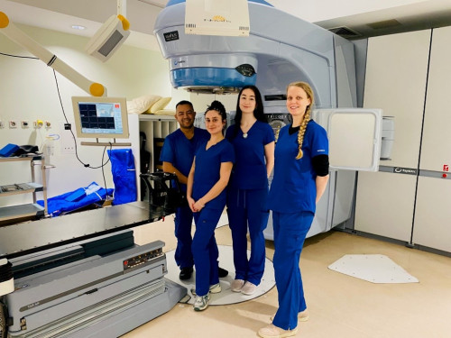
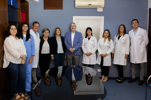
Warning: Undefined array key "category" in /var/www/novaliscircle.org/current/html/modules/aqb/articles/classes/AqbArticlesSearchResult.php on line 86 User Stories by RSSUser Stories by RSSTue, 04 Aug 2026 09:42:06 GMT<![CDATA[Radiologie München advances breast cancer treatments with prone deep inspiration breath hold and ExacTrac Dynamic Surface]]>Radiologie München, a private healthcare group, operates three radiotherapy sites across Munich, Germany with a total of five linear accelerators, one with ExacTrac Dynamic and three with ExacTrac Dynamic Surface. Treating approximately 1,500 tumor patients annually, their focus lies on improving treatment quality and the patient experience while also increasing efficiency.Following the installation of Brainlab ExacTrac Dynamic Surface in May 2025 on an Elekta Versa HD linac, the device has been fully integrated into their routine clinical workflows. It is utilized for initial patient setup and intrafraction monitoring for all patients and indications, including deep inspiration breath hold (DIBH) treatments for breast cancer patients.Dr. Gregor Habl, radiation oncologist at Radiologie München, shared insights gained from patients and their clinical team: “The ability to perform treatments supported by ExacTrac Dynamic Surface without permanent skin markings has contributed to improved patient comfort, while also streamlining workflows and enabling treatment durations of approximately 10 minutes per session.”Exploring the benefits of prone DIBHThe implementation of prone positioning in combination with DIBH has recently been expanded for breast cancer treatments at Radiologie München. The technique was introduced and established in the clinic by the clinical team and their medical physicist colleague Dr. Nils Temme, who brought his clinical experience with a prone breast board from his previous institution.Dr. Steffen Hennies, radiation oncologist at Radiologie München, explained: “This approach provides significant dosimetric advantages, particularly for left-sided breast cancer, by further reducing radiation exposure to organs at risk, including the heart and lung. While prone positioning offers substantial benefits, one challenge is that gravity can cause the heart to move closer to the anterior thoracic wall and therefore nearer to the breast target volume. DIBH... Read more]]>Tue, 04 Aug 2026 09:42:06 GMT<![CDATA[Angio excellence Down Under: Elements Image Fusion Angio and SmartBrush® Angio powering frameless AVM radiosurgery in Australia]]>This month, we are proud to celebrate the radiosurgery team at Prince of Wales Hospital in Randwick, Australia, for transforming the arteriovenous malformation (AVM) care they provide. They’ve done this by adopting frameless radiosurgery with digital subtraction angiography (DSA), enabled by Brainlab Elements Image Fusion Angio and Elements SmartBrush Angio.Having now treated over 100 AVM patients using this dedicated workflow, the team recognizes both the departmental impact and scalability of this innovative approach in routine practice.The challenge: Complex targets, logistical constraints. An AVM is an abnormal tangle of blood vessels for which radiosurgery can be a treatment option for select patients. Historically, AVM radiosurgery has relied on rigid immobilization with stereotactic head frames for accurate patient positioning and co-registration of multiple imaging studies, including 2D DSA and 3D image scans.This traditional, frame-based process can lead to:Patient discomfort from invasive frame placement and wearing a frame throughout a full treatment dayLogistical demands for the clinical team to schedule different imaging modalities with subsequent treatment planning and delivery the same dayRepeat invasive angiography for treatment planning purposesThe innovation: Frameless treatments for AVM that incorporate DSA imaging. In a first for Australia, the team implemented Brainlab Elements solutions for angiography/DSA, a frameless approach for planning AVM treatments. With Elements Image Fusion Angio, multiple 2D DSA images can be co-registered using cerebrovascular structures with 3D planning CT or MR images. Clinicians can thereby simultaneously outline vascular targets in 2D and 3D datasets. The software also supports diagnostic 2D DSA images without requiring an additional frame-based angiography procedure.This workflow enables:AVM contouring using multiple imaging modalities, including 2D DSA to improve visualization and spatial resolution of the nidu... Read more]]>Tue, 09 Jun 2026 15:10:18 GMT<![CDATA[The largest craniospinal radiosurgery service in Ireland]]>St. Luke’s Radiation Oncology Network: Leveraging Brainlab advanced stereotactic planning and image-guided patient positioning and monitoring to support brain and spine radiosurgery.As Ireland’s largest public radiation oncology provider, St. Luke’s Radiation Oncology Network operates across three sites in Dublin, Ireland. Two of the centers are located on the campuses of major acute hospitals: St. James and Beaumont. The Beaumont Hospital site serves as the national referral center for craniospinal and skull base radiosurgery treatment, delivering specialized care for complex intracranial and spinal conditions.The challenge: Achieving the seemingly unachievable. With rising demand for both intracranial and spine radiosurgery, the Beaumont RCSI Cancer Centre team needs to deliver precise, consistent treatments in both routine and high-risk scenarios while maintaining efficiency across a high-volume clinical schedule. This becomes especially critical in spine radiosurgery, where clinicians aim to deliver a sufficiently high dose to control the tumor while sparing the spinal cord just millimeters away. Achieving this balance requires a highly advanced algorithm—one that can sculpt dose in a concave target such as the vertebrae and create high-gradient plans that protect the cord without compromising tumor coverage. Once the optimal plan is created, the patient needs to be correctly positioned and monitored throughout treatment to enable intended delivery, with submillimetric accuracy from start to finish if needed. Multidisciplinary collaboration and robust high-quality technology are essential for this busy center that treats many different indications, from purely palliative to extremely complex radiosurgery.The innovation: “Brainlab technology is deeply integrated into Beaumont RCSI Cancer Centre’s radiosurgery department.” Central to the Network’s approach are Elements Radiosurgery Solutions and ExacTrac, a configuration that supports complex radiotherapy planning... Read more]]>Tue, 05 May 2026 09:09:13 GMT<![CDATA[Leading by example through knowledge sharing and Novalis Certification]]>For more than a decade, Instituto Zunino in Córdoba, Argentina has been recognized as a regional reference center for high-precision radiotherapy. Built on long-standing clinical expertise and a strong culture of quality, the center has developed a mature SRS and SBRT program that continues to evolve through structured processes, external validation and a commitment to shared learning.The team’s journey with Brainlab technology began in 2009 with iPlan RT and expanded in 2011 with ExacTrac X-Ray. Today, RT Elements is clinically adopted across the department of four treatment machines, supported by ExacTrac Dynamic—forming the technical backbone of daily practice.A benchmark for quality: Novalis CertificationAt Instituto Zunino, maintaining excellence means welcoming independent evaluation. First Novalis Certified in 2016, and successfully re-certified in 2020 and 2025, the team has consistently demonstrated its commitment to quality, safety and transparency. The most recent re-certification reinforces the center’s alignment with international standards and its place within the Novalis Certified community.Why certification matters—in their own wordsSilvia Zunino, MD, PhD, Director and Founder of Instituto Zunino and Fundación Marie Curie, explains: “Novalis Certification confirms that we work according to the same standards as leading centers in the United States and Europe. It gives us confidence in our methods and supports continuous improvement.”Daniel Venencia, PhD, Head of Medical Physics, adds: “Even as an experienced institution, having our work audited helps us identify opportunities to improve. Clearly defined protocols and external review ensure our patients receive the best possible treatment.” Motivated by his commitment to excellence in SRS and SBRT and to support his colleagues, Daniel Venencia has now joined the Novalis Certification Program team as an auditor.Beyond the clinic: sharing expertise to advance the regionInstituto Zunino’s impact extends ... Read more]]>Tue, 10 Mar 2026 09:18:17 GMT
 ]]>
]]> ]]>
]]> ]]>
]]> ]]>
]]>