


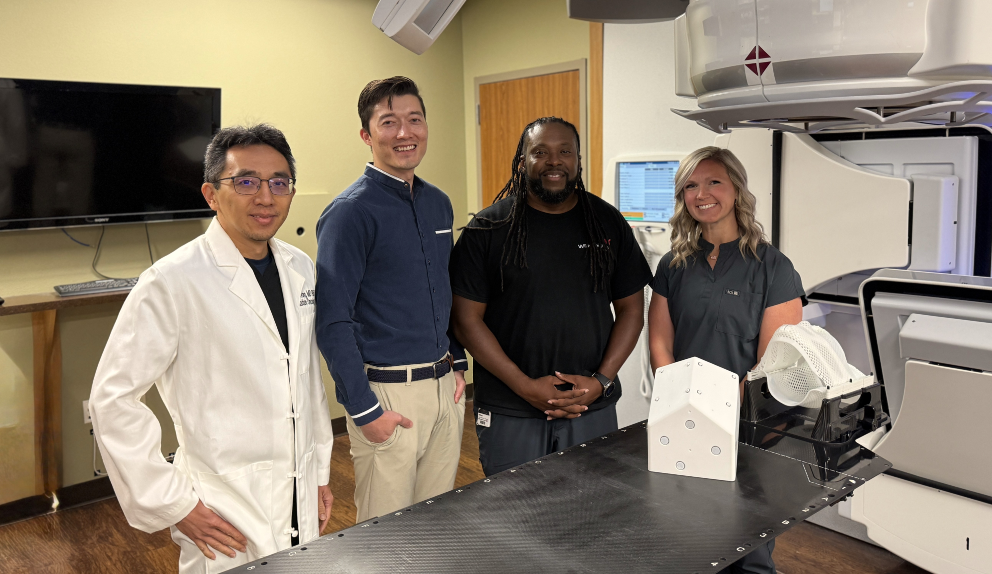
The Department of Radiation Oncology at Willis Knighton Cancer Center in Shreveport, Louisiana, USA opened in 1997. Since then, innovation has been at the core of their community oncology practice. The department became one of the first in the nation to offer solutions now regarded as standard, such as image-guided radiation therapy (IGRT) and intensity-modulated radiation therapy (IMRT). They continued building on this legacy, becoming the first site in the world to deliver stereotactic body radiation therapy (SBRT) for lung tumors on a helical delivery system, installing the world’s first commercial compact pencil beam scanning proton therapy system in 2014, and most recently, launching their Nuclear Oncology Division dedicated to radiotheranostics.
The challenge: keeping patients close to home
Before Willis Knighton Cancer Center started with these dedicated programs, patients in the Shreveport area often had to travel out of state, sometimes three to four hours each way, to access specialized radiosurgery, placing a significant burden on them and their families.
The response: building a robust SRS program in state
To keep treatment closer to home for even more patients, Willis Knighton Cancer Center expanded its services to include linac-based radiosurgery, giving patients in the local community access to advanced treatment capabilities. In 2016, Willis Knighton acquired Brainlab ExacTrac and Elements Multiple Brain Mets SRS. That investment marked the starting point of a radiosurgery program that has grown steadily in both volume and clinical scope.
The department went on to add further Brainlab Elements functional and vascular workflows, expanding its capabilities to treat not only patients with multiple brain metastases but also other indications such as trigeminal neuralgia and arteriovenous malformations (AVMs). Integrating these multi-modal contouring tools enabled them to work efficiently across the different specialties, including nuclear oncology and radiology.
Today, the team performs over 50 stereotactic radiosurgery (SRS) cases every year, using Brainlab Elements radiosurgery solutions for treatment planning and ExacTrac for precise patient positioning and monitoring across their two treatment machines.
As the team treated more and more patients with multiple brain metastases, retreatment became an important part of clinical practice. To support these increasingly complex cases, Willis Knighton Cancer Center added Brainlab Elements Retreatment Review to assess previous and planned treatments.
“Having intuitive dose summation tools like Elements Retreatment Review helps guide our treatment planning to ensure that we can manage these challenging cases optimally.” - Yohan Walter, MS, DABR, DABSNM, SRS Physics Lead, Chief of Radiation Physics Research, and Director of Nuclear Oncology Physics at Willis Knighton Cancer Center
The impact: rapid, precise and closer to home
The most tangible result is access. Willis Knighton Cancer Center is now delivering rapid, precise treatment across the full range of cranial indications using Brainlab solutions. Complex cases that were previously out of scope, like trigeminal neuralgia and AVMs, are now routine for the center.

Figure: Heat map illustrating the geographic reach of the Willis Knighton radiosurgery program: nearly 60% of patients live within 20 miles (32 km) of the cancer center.
In their words: “When we started this program in 2016, our goal was keeping patients in state for the best-in-class care they deserve. Today, with Brainlab technology guiding every step of planning and delivery, we're treating conditions we couldn't have taken on a few years ago, such as trigeminal neuralgia, AVMs or multiple brain metastases with the same precision you'd find at any bigger center. What means the most to me is seeing patients go home the same day, close to their families, instead of managing a diagnosis hundreds of miles from everything familiar to them.” - Chiachien Jake Wang, MD, PhD, Radiation Oncologist and Clinical Chief of Nuclear Oncology at Willis Knighton Cancer Center
Why it matters
Willis Knighton Cancer Center has built a respected regional radiosurgery program, having treated over 300 SRS patients across a broad range of conditions.
The statements made by the clinicians represent their personal opinions and experiences. These statements may not be supported by scientific evidence or peer-reviewed research. For verified information about any device mentioned, please refer to the manufacturer's official documentation.

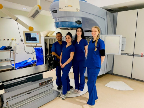
St. Luke’s Radiation Oncology Network: Leveraging Brainlab advanced stereotactic planning and image-guided patient positioning and monitoring to support brain and spine radiosurgery.
As Ireland’s largest public radiation oncology provider, St. Luke’s Radiation Oncology Network operates across three sites in Dublin, Ireland. Two of the centers are located on the campuses of major acute hospitals: St. James and Beaumont. The Beaumont Hospital site serves as the national referral center for craniospinal and skull base radiosurgery treatment, delivering specialized care for complex intracranial and spinal conditions.
The challenge: Achieving the seemingly unachievable. With rising demand for both intracranial and spine radiosurgery, the Beaumont RCSI Cancer Centre team needs to deliver precise, consistent treatments in both routine and high-risk scenarios while maintaining efficiency across a high-volume clinical schedule. This becomes especially critical in spine radiosurgery, where clinicians aim to deliver a sufficiently high dose to control the tumor while sparing the spinal cord just millimeters away. Achieving this balance requires a highly advanced algorithm—one that can sculpt dose in a concave target such as the vertebrae and create high-gradient plans that protect the cord without compromising tumor coverage. Once the optimal plan is created, the patient needs to be correctly positioned and monitored throughout treatment to enable intended delivery, with submillimetric accuracy from start to finish if needed. Multidisciplinary collaboration and robust high-quality technology are essential for this busy center that treats many different indications, from purely palliative to extremely complex radiosurgery.
The innovation: “Brainlab technology is deeply integrated into Beaumont RCSI Cancer Centre’s radiosurgery department.” Central to the Network’s approach are Elements Radiosurgery Solutions and ExacTrac, a configuration that supports complex radiotherapy planning, precise patient positioning and monitoring for a wide range of conditions from spinal metastases to intracranial metastases and benign intracranial conditions (including arteriovenous malformations, or AVMs, vestibular schwannomas and trigeminal neuralgia).
The intracranial radiosurgery program began using Brainlab solutions in 2013 and Beaumont RCSI Cancer Centre was the first in Europe to offer frameless radiosurgery treatments for trigeminal neuralgia. Brainlab Elements was introduced in 2018, initially used to treat multiple brain metastases, and became the primary planning platform for central nervous system cases in 2022. Single isocenter planning for multiple brain lesions enabled more consistent scheduling and workflow efficiency for a busy center treating a wide range of conditions.
The Beaumont RCSI Cancer Centre features the St. Luke’s Network’s flagship linac, Delvin, named after a river in Ireland through a patient-led naming initiative. This linac is equipped with ExacTrac which through image guidance allows for reduction in setup margins, making it very attractive to all consultants.
There is constant high demand for Delvin treatment slots and the ability to treat multiple brain metastases with a single isocenter has been practice changing for the Network. Today, nearly a third of the Network’s metastatic brain patients are treated using Elements Multiple Brain Mets SRS with a single isocenter.
Elements Radiosurgery Solutions have become a cornerstone of the Network’s rapidly expanding spine radiosurgery program. Since patients cannot be in the same position for their MR and CT scans, Elements Curvature Correction Spine—a dedicated deformable co-registration tool—enables them to use information from both modalities to better define the spinal anatomy and targets. In combination with AI-powered extra-cranial segmentation, organs at risk can be easily segmented.
In their words: “Our spine radiosurgery service has taken off over the last couple of years. We have designed a country-wide clinical trial to evaluate the safe dose escalation for patients with spinal metastases which is reaching our accrual targets much faster than expected.” — Professor Clare Faul, Consultant Radiation Oncologist at St. Luke’s Radiation Oncology Network.
Plans are generated using Elements Spine SRS, which enables accurate dose plan optimization and calculation with a Monte Carlo algorithm, which the team considers the most significant gain.
“All two-fraction patients are planned exclusively in Elements Spine SRS. Internal plan comparisons show a far better conformity to the target and sparing of organs at risk for those plans created in RT Elements. Possibly the most significant gain is the ability to optimize and calculate with a true Monte Carlo algorithm. With the newest software release, the fast Monte Carlo allows us to optimize and calculate with high accuracy in less than 20 minutes. The dedicated optimizer is extraordinary, too. We have been able to dose escalate in high-risk patients, where over 50% of the target is made of the gross target volume (GTV), without compromising proximal organs at risk. We get very excited every time we achieve the seemingly unachievable.” — Dr. Christina Skourou, Senior Physicist at St. Luke’s Radiation Oncology Network.
Treating so close to the spinal cord, nerve roots and esophagus are inherently challenging. For this reason, early on, the team performed extensive setup validation to establish safe and realistic planning target volume (PTV) margins. They would also acquire multiple CBCTs during each treatment fraction, but as they gained confidence in ExacTrac, they realized they could safely reduce patient imaging dose and shorten treatment time by relying on the device alone for intrafraction monitoring and correction. Today, spine stereotactic ablative radiotherapy (SABR) patients receive one posterior kV image to confirm the correct vertebral level, a CBCT to verify internal anatomy against plan and multiple ExacTrac kV images throughout treatment. Overall, treatment time per fraction is only slightly longer than for conventionally fractionated treatments.
The impact: Expanding access to radiosurgery for patients with complex brain and spine conditions. Together, Elements Radiosurgery Solutions and ExacTrac offer the Beaumont team greater confidence to deliver radiosurgery in high-risk scenarios. Consistent target definition and high-quality dose planning, together with precise patient positioning and continuous monitoring, enable submillimetric accuracy throughout treatment. The high level of accuracy supports evolving treatment pathways and allows radiosurgery to be integrated into clinical decision-making across a wider array of conditions.
St Luke’s Radiation Oncology Network was the first center in Ireland to achieve Novalis Certification, underscoring its leadership in advanced stereotactic radiotherapy. “The Novalis Certified program has played an important role in quality assurance and patient safety at the Network by independently validating that stereotactic radiotherapy is delivered to the highest international standards” — Paul Davenport, Principal Physicist at St. Luke’s Radiation Oncology Network.
Pictures: © St. Luke’s Radiation Oncology Network, Dublin, Ireland
Disclaimer: The statements made by the clinicians represent their personal opinion and experience. These statements may not be supported by scientific evidence or peer-reviewed research. For verified information about the device, please refer to the manufacturer's official documentation.
The challenge: serving a large region with growing clinical complexity
In northern Poland, Centrum Onkologii im. prof. Franciszka Łukaszczyka w Bydgoszczy caters to a vast geographical region where many patients travel long distances for specialized care. As a center with extensive experience in treating tumors of the central nervous system, the demand for their services expanded, and the team faced a growing number of cases requiring high-quality radiosurgery.
Table 1: The number of SRS and SBRT treatments at the Oncology Center in Bydgoszczy has increased by almost 2,000% in less than a decade.
In 2024 alone, the team treated 340 patients with brain metastases, 71 with spinal metastases, 134 patients with meningiomas and 56 with vestibular schwannomas (VIII nerve)—plus over 350 extracranial cases.
Over more than 15 years, the center further developed their comprehensive SRS and SBRT program. What began in 2009 with BrainSCAN, the first treatment planning solution from Brainlab, continued through iPlan RT and has since evolved into the broad adoption of RT Elements across eight treatment machines, alongside Brainlab ExacTrac Dynamic. Increasing patient volumes as well as multifocal disease presentations pushed the team to refine workflows capable of handling high case complexity on a day-to-day basis.
As case numbers and complexity continued to rise, the team began to push conventional planning boundaries—not just in theory, but in daily clinical practice. One case treated in May 2025 became a clear example of how their approach could scale even to highly extensive disease presentations.
The case: planning for 34 brain metastases in a single patient
A 67-year-old man presented with headaches, nausea and vomiting. Imaging revealed 34 brain metastases along with an adrenal lesion associated with newly diagnosed lung adenocarcinoma. Despite the high lesion count, the patient’s clinical condition remained good, and systemic therapy offered a meaningful survival outlook. The team elected to pursue SRS for all intracranial lesions—an approach once considered impractical at this scale.
Using Brainlab Elements Multiple Brain Mets SRS, the clinicians were able to treat all lesions using only two isocenters during the same planning session:
- Isocenter 1: 10 targets
- Isocenter 2: 24 targets
- Total GTV: 18.6 cm³
- Total PTV: 26 cm³
- Dose: 25 Gy in 5 fractions
While highly complex, the case aligned with the department’s established planning and treatment workflow.
The impact: a strong clinical response at first follow-up
At the initial follow-up in September, the results were striking:
- 33 lesions demonstrated complete regression
- 1 lesion showed partial regression
- the patient reported no neurological deficits
- no steroid therapy was required
- and he remained asymptomatic
For the Bydgoszcz team, the case underscored a pattern they had observed across growing patient volumes: when planning tools support structured segmentation and a monoisocentric approach, lesion count becomes less of a technical barrier to delivering treatment.
Prof. Maciej Harat, MD, and Maciej Blok, PhD, MD, Centrum Onkologii Im. Prof. Franciszka Łukaszczyka W Bydgoszczy reflect on the experience: “This case showed us that the number of brain metastases is no longer the limiting factor for delivering focal treatments. Alongside these highly complex treatments, we are managing an increasing number of patients each year—and we would not be able to keep pace without Brainlab Elements. The software supports automatic segmentation of targets and organs at risk and helps us generate highly complex plans in a very short time.”
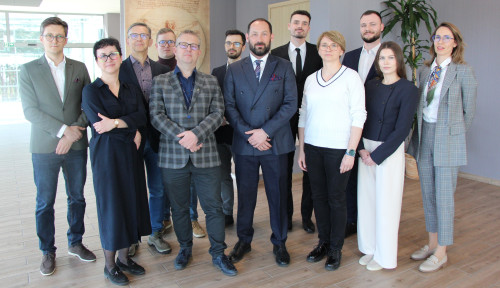
The team behind the progress: 25 radiation oncologists, 26 medical physicists, 38 radiation therapists and more clinical staff at Bydgoszcz have built a radiosurgery program capable of treating some of the region’s most complex cases. Their commitment ensures that patients across a large geographical area can access advanced, high-quality care.
Please join us in celebrating the team at Centrum Onkologii im. prof. Franciszka Łukaszczyka w Bydgoszczy for demonstrating how skills and well-established workflows can expand access to precise radiosurgery—no matter the complexity.
Picture: ©Centrum Onkologii im. prof. Franciszka Łukaszczyka w Bydgoszczy, Poland
The statements made by the clinicians represent their personal opinion and experience. These statements may not be supported by scientific evidence or peer-reviewed research. For verified information about the device, please refer to the manufacturer's official documentation.

From a clinical workflow and technology standpoint, I was wondering—given the geometric limitations in ring gantry systems, is the likely direction toward surface imaging with AI-based prediction, or are there ongoing developments for internal tracking within closed-bore architectures?
First of all thank you for this platform.
May i know Sir from your experience, what are the most effective strategies for managing intrafraction motion during SBRT, particularly in spine and lung cases?



Join us in Philadelphia for our Mid-Atlantic Regional Symposium with insightful clinical talks by renowned clinical experts and inspirational discussions on SRS and SBRT.
Experience cutting edge of radiotherapy and radiosurgery at our exciting two days symposium focused on precision, personalization, and standardization in Stereotactic Radiosurgery (SRS) and Stereotactic Body Radiation Therapy (SBRT).
This event will bring together medical physicists, dosimetrists, radiation oncologists and radiation therapists to share valuable clinical techniques and innovative management strategies, as well as to gain insights into how complex data is reshaping the field of radiosurgery.
Brainlab Mid-Atlantic Regional Symposium
May 8 and 9, 2026
Click here to have the program.
We look forward to welcoming you to Philadelphia for this inspiring event!
To register click here

At TUM University Hospital in Munich, the radiotherapy team faces a growing number of complex cranial cases. From patients with dozens of brain metastases to those requiring retreatment, manual contouring can be time-intensive and variable across operators. As case volumes rise, the team has been exploring new ways to maintain consistency and efficiency without compromising clinical accuracy.
The challenge: segmentation at scale
For radiosurgery teams, delineating multiple metastases or large primary tumors is one of the most demanding steps in planning. Each contour must be precise, consistent and reproducible. As the number of lesions increases, so does the time required for manual work and the risk of inter-operator variability. The team at TUM University Hospital needed a way to standardize segmentation while keeping pace with growing treatment demands.
The innovation: AI-powered segmentation integrated into routine workflows
TUM University Hospital became one of the first clinical centers to implement Brainlab Elements AI Tumor Segmentation in daily practice. Utilizing contrast-enhanced T1-weighted MRI scans, the software automatically identifies enhancing tumor tissue using a neural-network-based model trained on more than 4,000 scans and 11,000 expert annotations.
The workflow supports:
- Semi-automatic segmentation of multiple brain metastases for SRS
- Semi-automatic segmentation of other cranial tumors, including meningiomas, cranial nerve tumors, gliomas, glioneuronal and neuronal tumors
- Fast processing with a segmentation duration of under one minute per contrast-enhanced T1-weighted MR image set*
Within just one month, the team incorporated the AI tool into all cranial planning workflows, including Gamma Knife (Elekta AB, Sweden) and retreatment reviews.
The shift: efficiency that reinforces precision
Early observations at TUM University Hospital show strong performance for brain metastases larger than 3 mm and primary tumors over 10 mm. By automating the most time-consuming parts of segmentation, the team can focus more attention on clinical decision-making and the nuances of each case. As one clinician noted, the practical impact becomes clear when treating patients with numerous lesions. Elements AI Tumor Segmentation helps standardize the foundation of the plan, allowing experts to build on it with confidence.
In their words: “Using Elements AI Tumor Segmentation enhances our multiple brain metastases and retreatment review workflows. We are very happy with the results after implementing this innovative AI method in our clinic.” — Jan Peeken, MD, PhD, Senior Managing Radiation Oncologist at TUM University Hospital.
The impact: AI as a meaningful part of clinical routine
For a center treating complex cranial cases at high volume, automated segmentation is becoming an important tool that supports consistency and efficiency across the radiosurgery workflow. By integrating Elements AI Tumor Segmentation directly into daily practice, the team is shaping a future where technology strengthens—not replaces—clinical expertise.
Please join us in recognizing TUM University Hospital for helping pave the way for practical, clinically grounded AI adoption in cranial radiosurgery.
* Valid in a widely available platform like Brainlab Planning Station 9.0.1 or higher.
The statements made by the clinician represent their personal opinion and experience. These statements may not be supported by scientific evidence or peer-reviewed research. For verified information about the device, please refer to the manufacturer's official documentation.